FIGURE 7.
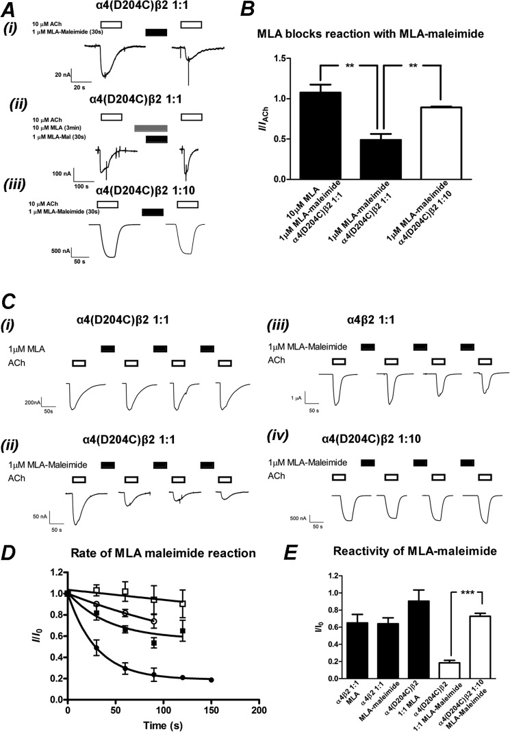
MLA-maleimide irreversibly inhibits α4(D204C)β2 nAChRs injected in a 1:1 but not 1:10 ratio. A, shown is the initial trapping experiment of MLA-maleimide to the introduced cysteine. i, 10 μm ACh was applied to α4(D204C)β2 nAChRs injected in a 1:1 ratio (open bar). 1 μm MLA-maleimide was applied for 30 s followed by a 17-min wash with buffer to remove excess MLA-maleimide. 10 μm ACh was applied again, demonstrating an irreversible reduction in the ACh-elicited response. ii, shown is an identical experiment, except 10 μm MLA were incubated for 3 min then co-applied with 1 μm MLA-maleimide followed by a 17-min wash and application of 10 μm ACh. iii, 1 μm ACh was applied to α4(D204C)β2 nAChRs injected in a 1:10 ratio (open bar). 1 μm MLA-maleimide was applied for 30 s followed by a 17-min wash with buffer to remove excess MLA-maleimide. 1 μm ACh was applied again, blocking the irreversible reduction in the ACh-elicited response. B, shown is the mean ± S.E. of ACh-elicited responses after 30 s of treatment of 1 μm MLA-maleimide and a 17-min wash, normalized to the initial ACh-elicited response. 1 μm MLA-maleimide significantly reduced the ACh-elicited response in α4(D204C)β2 nAChRs injected in a 1:1 ratio (filled bar, middle) compared with a 1:10 (open bar) ratio (p < 0.01, n = 3–4; Student's t test). Preincubation for 3 min and co-application of 10 μm MLA with 1 μm MLA-maleimide prevented the MLA-maleimide reaction (filled bars, left), significantly blocking the reduction of the ACh-elicited response at the α4(D204C)β2 nAChR injected in a 1:1 ratio (p < 0.01, n = 4–6; Student's t test). C, rate of reaction experiments with individual oocytes expressing α4(D204C)β2 nAChRs injected in a 1:1 ratio (i and ii), α4β2 nAChRs injected in a 1:1 ratio (iii), and α4(D204C)β2 nAChRs injected in a 1:10 ratio (iv). An EC50 concentration of ACh (10, 10, 100, and 1 μm for (i–iv), open bars) was applied before application of 1 μm MLA (i) or) MLA-maleimide (filled bars) (ii–iv. Each successive 30-s application of MLA or MLA-maleimide was followed by a 17-min wash with buffer to remove excess MLA and then an ACh application to measure the reduction in current resulting from trapping of the MLA-maleimide with the cysteine. D, shown is the mean ± S.E. of ACh normalized to the initial response to ACh after the cumulative time of successive 1 μm MLA or MLA-maleimide applications. Rates of reaction of MLA-maleimide to (α4[D204C])3(β2)2 (●), (α4[D204C])2(β2)3 (○), and α4β2 wild type (■) and MLA to (α4[D204C])3(β2)2 (□) are plotted with a single exponential fit to the average responses shown as described under “Experimental Procedures” (n = 3–6 for each rate of reaction). E, shown is the mean ± S.E. of the maximum reduction of ACh-elicited current as a fraction of the initial ACh-elicited current. The maximum reduction in current was determined either by fitting each individual experiment to a single exponential decay (α4(D204C)β2 1:1 and 1:10 ratio and MLA-maleimide; open bars) or the reduction in ACh-elicited response after 120 s of cumulative MLA or MLA-maleimide addition (α4β2 and MLA or MLA-maleimide and α4(D204C)β2 1:1 and MLA). The reduction in ACh-elicited response is significantly greater for α4D204Cβ2 injected in the 1:1 compared with the 1:10 ratio (p < 0.001, n = 3- 5), indicating that MLA-maleimide is being trapped at the D204C residue in the α4-α4 interface (white bars). The ACh-response recovers up to 90% of the initial response, significantly different to the application of MLA-maleimide applied to the (α4[D204C])3(β2)2 nAChR, injected in a 1:1 ratio (p < 0.01, n ≥ 3; Student's t test).

