FIGURE 7.
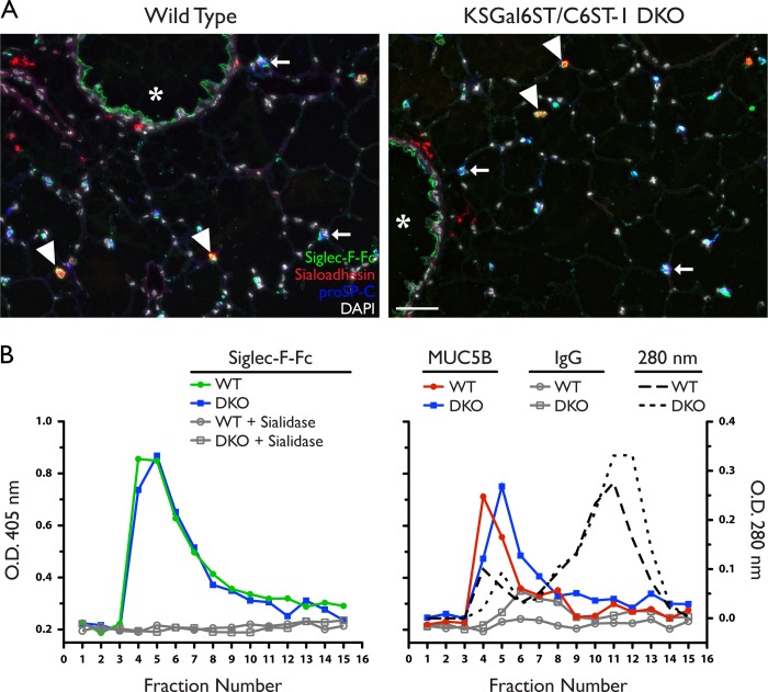
Siglec-F ligand expression in lungs from KSGal6ST/C6ST-1 DKO mice. A, cryostat-cut sections of lungs from WT or DKO mice stained with Siglec-F-Fc (green), anti-sialoadhesin (red), anti-proSP-C (blue), and DAPI (white). Scale bar represents 50 μm. Results are representative of two independent experiments. B, Siglec-F-Fc reactivity (left) in BAL fluid fractions from WT (filled circles) or DKO mice (filled squares), assayed by ELISA. Results are representative of two independent experiments. The signal was eliminated by sialidase treatment (open circles, open squares). Anti-MUC5B reactivity (right) in BAL fluid fractions from WT (filled circles) and DKO mice (filled squares) was assayed by ELISA. Isotype control signal was minimal (open circles, open squares). Total protein was determined by measuring absorbance at 280 nm for wild type (dotted line) and DKO mice (dashed line). WT, wild type; DKO, KSGal6ST/C6ST-1 double knock-out.

