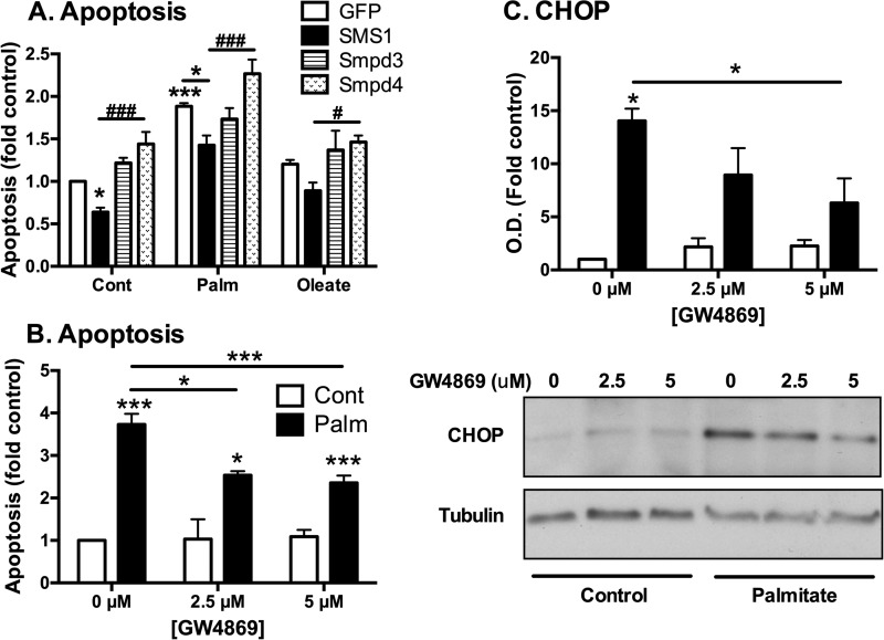
FIGURE 6.
SM content and SMase activity play a significant role in palmitate-induced ER stress and apoptosis. A, MIN6 cells were transfected with GFP, SMS1, Smpd3, or Smpd4 constructs prior to 48-h palmitate (Palm) or oleate (0.4 mm/0.92% BSA) treatment and then quantified for the level of apoptosis. #, p < 0.05; ###, p < 0.001; two-way analysis of variance with Bonferroni's multiple comparisons of Smpd4 to SMS1 where indicated; *, p < 0.05; ***, p < 0.001; unpaired Student's t tests compared with GFP control unless indicated. Data represent mean apoptosis (fold control) + S.E. from three to four independent experiments. Cont, control. B, MIN6 cells were cultured with 0.4 mm palmitate complexed to BSA (0.92%) ± SMase inhibitor, GW4869, for 48 h before total lysates were prepared for the quantification of apoptosis (DNA fragmentation via cell death ELISA, Roche) or CHOP induction (immunoblotting) (C). *, p < 0.05; **, p < 0.01; ***, p < 0.001; two-way analysis of variance with Bonferroni's multiple comparisons to control (0 μm) or where indicated.

