
FIGURE 6.
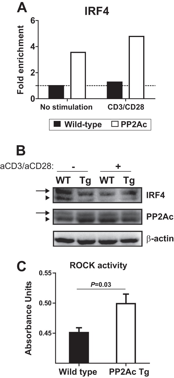
PP2Ac promotes IRF4 activation and recruitment to the Il17a locus. A, ChIP experiments were performed to analyze IRF4 occupancy of the Il17a promoter region in naïve CD4 T cells before and after stimulation with anti-CD3 and anti-CD28. Results were normalized against the input and are expressed as fold change over the unstimulated WT cells. B, naïve CD4 T cells were isolated from PP2Ac Tg and WT mice and were lysed before or after activation with anti-CD3 and anti-CD28 (1 h). Lysates were separated in a SuperSep Phos-tag acrylamide gel that retards the migration of phosphorylated proteins. Proteins were transferred to a PVDF membrane that was blotted with anti-IRF4, anti-PP2Ac, and anti-β-actin. Arrows indicate the phosphorylated proteins. Arrowheads indicate unphosphorylated proteins. C, cell lysates of stimulated naïve CD4 T cells were probed for Rho kinase activity.