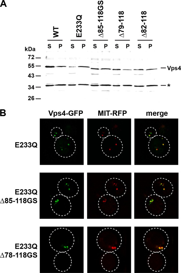
FIGURE 2.

Subcellular localization of mutant Vps4 proteins. A, Western blot analysis of soluble (S) and membrane-bound pellet (P) cell fractions using anti-Vps4 antiserum. The cross-reacting protein at 35 kDa (*) serves as a loading control. B, fluorescence microscopy of cells expressing GFP-tagged versions of Vps4 mutants and the MVB marker MIT-RFP.
