
FIGURE 5.
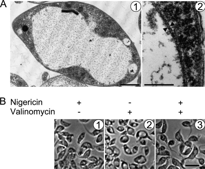
Treatment of PS T. brucei with the K+ ionophore valinomycin results in mitochondrial swelling. A, transmission electron micrographs of PS T. brucei treated with a 1 μm concentration of the K+ ionophore valinomycin. Double membranes are indicated by opposing arrowheads. Scale bars, 2 μm and 200 nm for pictures 1 and 2, respectively. Note the peripherally located kinetoplast DNA disk. B, light micrographs showing cells treated with either 2 μm nigericin or 1 μm valinomycin alone or pretreated with the former and then treated with the latter, as indicated at the top of the images from left to right. Scale bar, 10 μm.