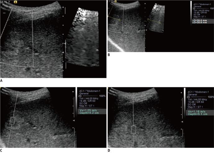
Fig. 3.
Hemangioma with same color and worse conspicuity on acoustic radiation force impulse (ARFI) 2-dimensional (2D) image of 55-year-old male.
A. This isoechoic hemangioma shows same stiffness as adjacent hepatic parenchyma on ARFI 2D image. Lesion conspicuity is worse on ARFI 2D image because of poor delineation of hyperechoic rim. B. Size of lesion is same on both images. C, D. Median shear wave velocity (SWV) of this hemangioma was 1.25 m/sec and mean SWV difference was 0.08 m/sec. Scales provided by dots in right vertical axis of B-mode images are in centimeters.

