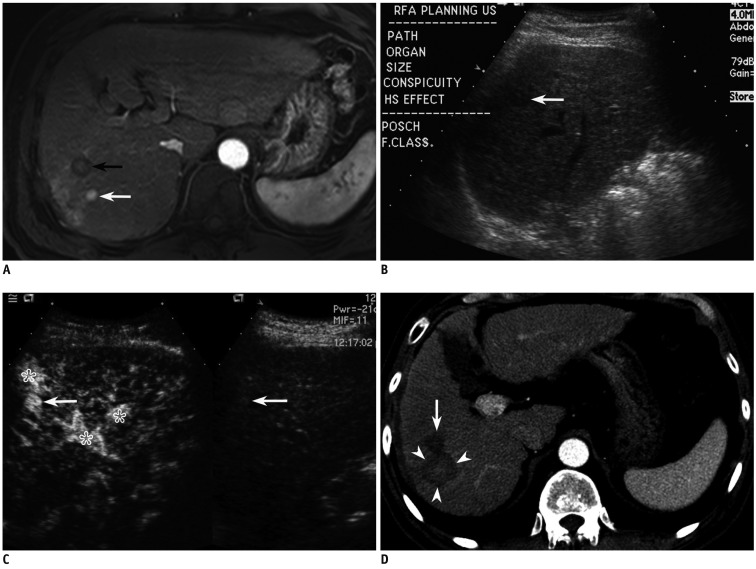
Fig. 3.
Fifty nine-year-old man with 1 cm-sized hepatocellular carcinoma (HCC) in segment VII of liver.
A. Arterial phase MR image (TR/TE, 3.1/1.5; flip angle, 10°; matrix size, 228 × 211; bandwidth, 724.1 Hz/pixel) shows 1.1 cm-sized enhancing HCC (white arrow) in segment VII of liver. Black arrow indicates previous radiofrequency ablation (RFA) zone. B. Small HCC candidate is seen as subtle low echoic nodule (arrow) with poor conspicuity in segment VII of liver on planning ultrasonography. C. On CEUS, which was performed to be certain, small enhancing nodule (arrow) was identified at same site as (B), suggestive of HCC. Asterisks indicate arterioportal shunts around index tumor. D. Arterial phase CT image obtained immediately after RFA shows large ablation zone (arrowheads) with sufficient ablative margin. Arrow indicates previous RFA zone.

