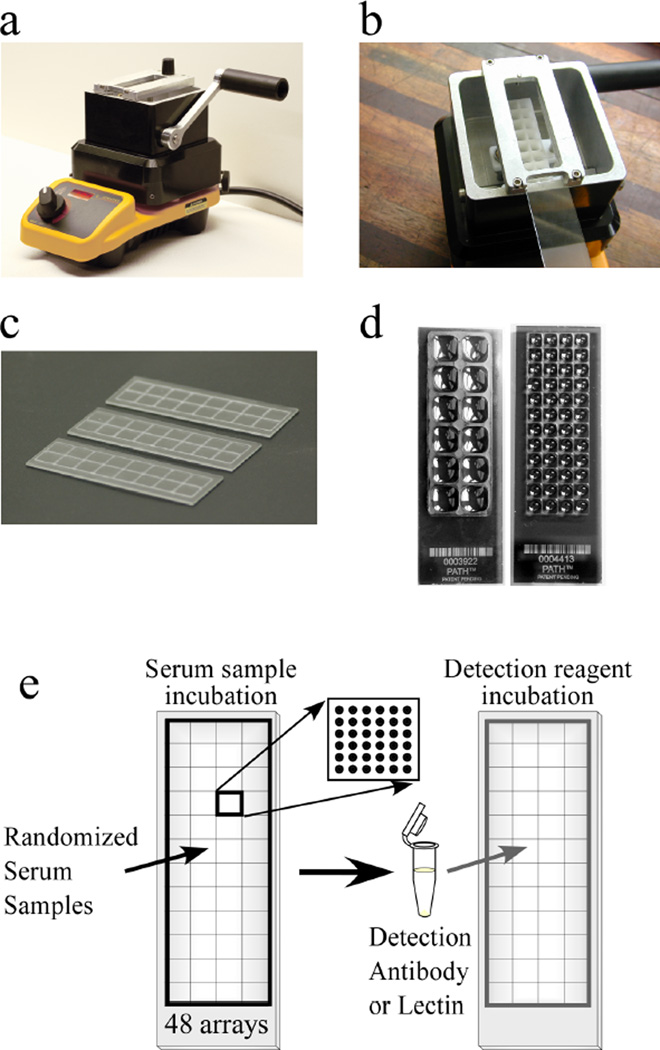
Figure 2.
High-throughput processing of antibody arrays. (a) A SlideImprinter device (The GelCompany, San Francisco, CA) is used to imprint wax patterns onto microscope slides. A bath of wax is melted on a hotplate, and (b) a slide is inserted upside-down into a holder above the bath. Pulling the level forward elevates a stamp out of the bath to contact the slide, (c) leaving a pattern of wax on the slide. (d) Various designs of stamps can be used, such as one that partitions 12 regions on a slide (left), or another that partitions 48 regions on a slide (right). The liquid loaded into each region remains segregated from the other regions. (e) Strategy for detecting a glycan structure on many samples. 48 different serum samples are randomized and incubated on 48 identical antibody arrays on a single microscope slide. Each array contains up to 144 spots. After the proteins bind to the antibodies, a glycan structure on the captured proteins are detected with a lectin or glycan-binding antibody.

