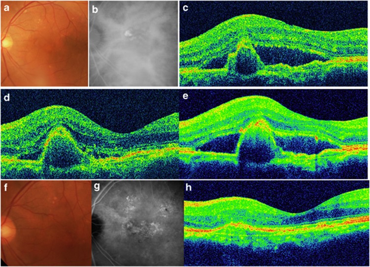
Figure 4.
A 75-year-old man presented with reduced visual acuity in the left eye. (a) A color fundus photograph of the left eye shows a large area of SRF. (b) Indocyanine green angiographic (ICGA) image showed staining indicating PCV. (c) Spectral-domain optical coherence tomography (SD-OCT) at baseline revealed SRF with a polypoidal lesion. The visual acuity was 20/30 in the left eye, and the patient was diagnosed as having PCV. (d) At 3 months after the first injection, the SRF had resolved, although the polypoidal lesion persisted. The patient's visual acuity had improved to 20/25. (e) However, at 13 months after the initial treatment, the SRF had increased. Additional IVR treatments were administered. (f) At 2 years after the first treatment, the patient's visual acuity was maintained at 20/25. (g) An ICGA image shows the regression of the polyps, although the abnormal vascular network remains. (h) SD-OCT also shows the disappearance of PCV. In total, 11 IVR treatments were administered during the follow-up period.

