
Fig. 6.
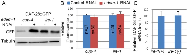
Misfolded DAF-28::GFP accumulates in ire-1 mutants. (A) Representative western blot of GFP and tubulin of coelomocyte-defective day-2 cup-4 and ire-1(ok799) mutants harboring an integrated DAF-28::GFP transgene. Animals were treated with control RNAi or edem-1 RNAi. edem-1 inactivation increased DAF-28::GFP levels in cup-4 mutants, but not in ire-1 mutants, whose basal DAF-28::GFP levels were high even before edem-1 inactivation. (B) Bar graph showing the relative mean whole-body fluorescence of DAF-28::GFP in day-2 cup-4 or ire-1 mutants treated with control RNAi (blue) or edem-1 RNAi (red). Fluorescence was calculated relative to control RNAi treatment of each strain. edem-1 inactivation did not increase DAF-28::GFP fluorescence. n, the number of animals analyzed. Similar results were obtained in two additional independent experiments. (C) Bar graph showing the average relative mRNA levels of daf-28::gfp in ire-1(+); cup-4(−) and ire-1(−) mutants, measured by qRT-PCR in three independent experiments. daf-28::gfp mRNA levels were not increased in ire-1 mutants (P>0.45, Student's t-test).