
Fig. 4.
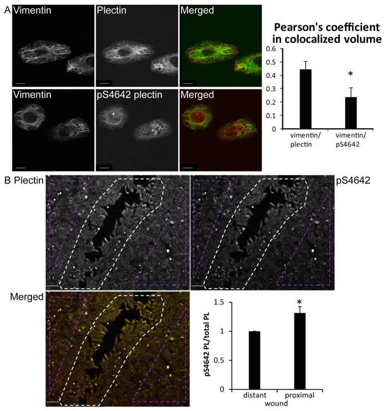
pS4642 plectin has an altered cytoplasmic distribution and increases during wound healing in SK-MEL-2 cells. (A) Confocal immunofluorescence microscopy images of SK-MEL-2 cells, immunostained with antibodies against plectin or pS4642 and vimentin as indicated. Degree of colocalization was determined by measuring the Pearson's coefficient using the Imaris software. Scale bar: 10 µm. More than 100 cells counted per slide. *P<0.01. (B) Confluent SK-MEL-2 cells were scratched, fixed 20 hours later and immunostained with anti-plectin and pS4642 antibodies. Scale bar: 100 µm. The ratio pS4642 plectin to plectin was determined in wound-proximal (white dotted area) and scratch-distant cells (violet dotted areas). Graphs show means ± s.d. *P<0.05 (n = 3).