
Fig. 5.
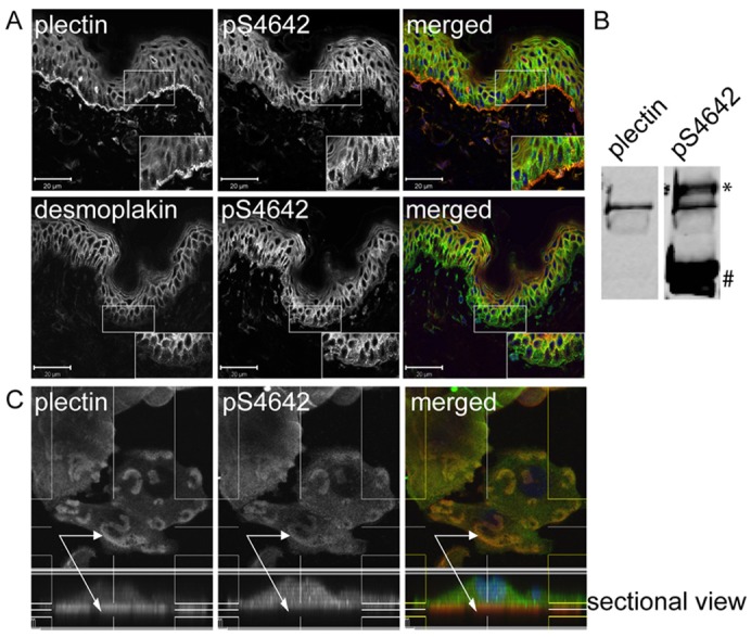
Plectin is less phosphorylated on S4642 at sites of cell-substrate contact in human epidermis and PAJEB-β4 keratinocytes. (A) Acetone-fixed cryosections of human skin were co-immunostained for plectin and pS4642 or for desmoplakin and pS4642. High-power views of the boxed areas are depicted in the insets. In the basal layer of the epidermis, there was no correlation between plectin and pS4642 signals whereas the immunostained patterns of desmoplakin and pS4642 were similar showing that plectin is weakly phosphorylated in hemidesmosomes. Scale bars: 20 µm. (B) Epidermal cell extracts were immunoblotted with anti-plectin and anti-pS4642 antibodies, indicating plectin S4642 phosphorylation in the skin (*, immunoreactive protein migrating above plectin; #, p2849 desmoplakin). (C) PAJEB-β4 keratinocytes, cultured in high Ca2+ for 24 hours, were fixed with formaldehyde and co-immunostained for plectin (red) and pS4642 (green) as indicated. Z-stack sectional view of plectin (red) and pS4642 (green) showing the dichotomy between the two signals in hemidesmosome-like structures (arrows indicate cell-substrate contact/hemidesmosome-like structures with a prevalence of unphosphorylated plectin in the lower part).