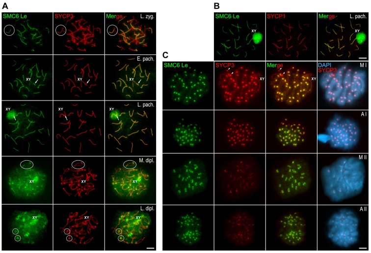
Fig. 2.
Distribution of SMC6 Le during meiosis. (A,C) Double-immunolabeling of SMC6 Le (green) and SYCP3 (red) on spread mouse spermatocytes at different meiotic stages: late zygotene (L. zyg.), early pachytene (E. pach.), late pachytene (L. pach.), mid diplotene (M. dipl.), late diplotene (L. dipl.), metaphase I (M I), anaphase I (A I), metaphase II (M II) and anaphase II (A II). Chromatin is stained with DAPI (blue) in C. Selected bivalents in late zygotene and mid diplotene panels, and centromere regions in the late diplotene panel, are encircled. The sex bivalent (XY) is indicated. Arrows in early pachytene and late pachytene panels indicate the PAR region between the sex chromosomes. Arrowheads in the metaphase I panel indicate large SYCP3 agglomerates in the cytoplasm. (B) Double-immunolabeling of SMC6 Le (green) and SYCP1 (red) on a spread late pachytene spermatocyte. The sex body (XY) is indicated. Scale bars: 10 µm.

