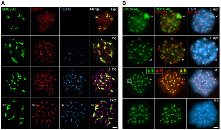
Fig. 3.
Distribution of SMC6 Ab during meiosis. (A) Triple-immunolabeling of SMC6 Ab (green), SYCP3 (red) and TEX12 (light blue) on spread leptotene (Lept.), early zygotene (E. zyg.), late zygotene (L. zyg.) and pachytene (Pach.) spermatocytes. The sex body (XY) is indicated in the pachytene panel. (B) Double-immunolabeling of SMC6 Ab (green) and SYCP3 (red) and counterstaining of the chromatin with DAPI (blue) on spread early diplotene (E. dipl.), late diplotene (L. dipl.), metaphase I (M I) and metaphase II (M II) spermatocytes. The sex body and the sex bivalent (XY) are indicated in the diplotene and metaphase I panels, respectively. The boxed area in the metaphase I panel is enlarged in the corresponding insets. Scale bars: 10 µm.

