
Fig. 4.
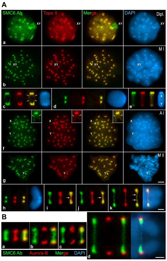
SMC6 Ab colocalizes with Topo IIα at prophase I chromocenters and at the centromeres during both meiotic divisions. (A) Double-immunolabeling of SMC6 Ab (green) and Topo IIα (red) and counterstaining of the chromatin with DAPI (blue) on spread diplotene (Dipl.), metaphase I (M I), anaphase I (A I) and metaphase II (M II) spermatocytes. Enlarged spread metaphase I autosomal (c,d) and sex bivalents (e), spread (h) and squashed (i, j) metaphase II chromosome, and squashed early anaphase II chromosome (k). The sex body (XY) in a, the sex bivalent (XY) in b and the sex chromosomes (X,Y) are indicated. The arrows in c and in the insets in f mark the chromatid axes, whereas in i–k, the arrows indicate a thin strand between the Topo IIα masses at the kinetochore regions. (B) Double-immunolabeling of SMC6 Ab (green) and Aurora-B (red) and counterstaining of the chromatin with DAPI (blue) on squashed metaphase II (a–c), and early anaphase II (d) spermatocytes. Scale bars: 10 µm (Aa,b,f,g); 5 µm (Ac–e,h–k and B).