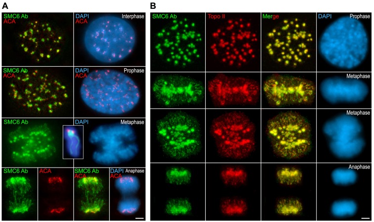
Fig. 5.
SMC6 Ab colocalizes with Topo IIα at the centromeres and chromatid axes in mitotic chromosomes. (A) Double-immunolabeling of SMC6 Ab (green) and an ACA serum revealing kinetochores (red) and counterstaining of the chromatin with DAPI (blue) on interphase, prophase, metaphase and anaphase 3T3 cultured cells. The inset in the metaphase panel shows an enlarged metaphase chromosome. (B) Double-immunolabeling of SMC6 Ab (green) and Topo IIα (red) and counterstaining of the chromatin with DAPI (blue) on prophase, metaphase and anaphase 3T3 cultured cells. Scale bars: 2.5 µm.

