
Fig. 2.
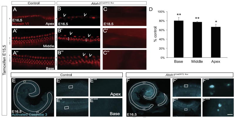
Cochlear hair cells die following Atoh1 deletion at E15.5. Pregnant dams received tamoxifen at E15.5. (A–C″) Cochleae from control littermate (A) and Atoh1CreERT2/flox (B–C″) embryos immunostained for myosin VI. (B–B″) E16.5 Atoh1CreERT2/flox mice lacked outer hair cells (1–3, brackets in B′) predominantly in the third row and scattered inner hair cells (I, arrowheads) throughout the cochlea. No hair cells were present at E18.5 (C–C″). (D) Myosin VI+ hair cell counts by region in Atoh1CreERT2/flox embryos as percent of control littermate numbers (n=3–4 cochleae/genotype). *p<0.05, **p<0.01 compared to control. (E–F″″) Activated caspase 3 immunostaining of cochleae from E16.5 embryos demonstrated apoptotic cell death in the organ of Corti (outlined) of Atoh1CreERT2/flox (F–F″″) but not littermate controls (E–E″″). Scale bar: 15 μm (A–C″), 500 μm (E, F); 50 μm (E′, E″, F′, and F″); 5 μm (E′″, E″″, F′″, and F″″).