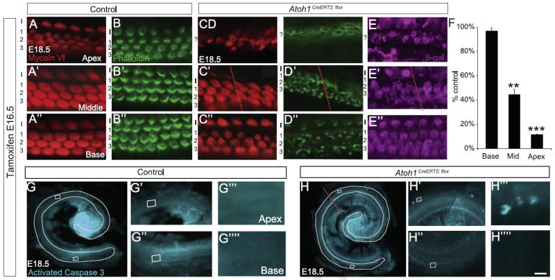
Fig. 3.

Atoh1 deletion at E16.5 causes hair cell loss and disrupts cochlear hair cell stereocilia formation. (A–D″) Cochleae from E18.5 littermate control (A–B″) and Atoh1CreERT2/flox (C–D″) embryos treated with tamoxifen at E16.5 and immunostained with myosin VI, β-galactosidase or labeled with phalloidin (A–A″, B–B″, C–C″, D–D″ and E–E″ are from different animals). Atoh1CreERT2/flox embryos have fewer hair cells from the mid-middle turn (red lines in C′, D′, E′ and H) to the apex while these cells are present in normal numbers from the mid-middle turn to the base. However, remaining hair cells have disorganized or no stereocilia (D–D″). Remaining hair cells also show evidence of recombination (E–E″). (F) Hair cell counts by cochlear region graphed as percent of control littermate numbers show significant loss of myosin VI+ hair cells in the apex and middle turn but not the base of Atoh1CreERT2/flox embryos (n=3 cochleae/genotype). **p<0.001, ***p<1 × 10⁻⁶. G–H″″) Immunostaining for activated caspase 3 shows cell death in the apex, but not the base, of Atoh1CreERT2/flox embryos. Scale bar: 15 μm (A–E″); 500 μm (G, H); 50 μm (G′, G″, H′, and H″); 5 μm (G′″, G″″, H′″, and H″″).
