Fig. 5.

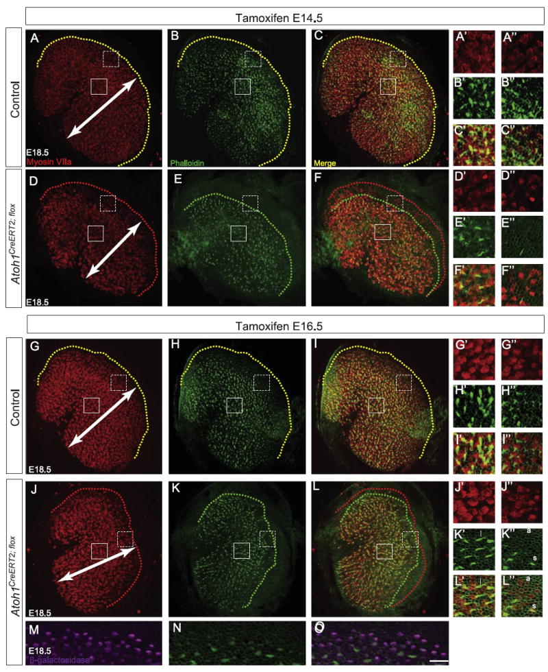
Atoh1CreERT2/flox embryos have fewer myosin VIIa+ cells and fewer stereociliary bundles in the utricle. Pregnant dams were gavaged with tamoxifen at either E14.5 (A–F″) or E16.5 (G–L″) and utricles harvested at E18.5. Immunostaining for myosin VIIa (A, D, G, and J) and labeling with phalloidin (B, E, H, and K) revealed decreased mediolateral extent (arrows) of the hair cell field in Atoh1CreERT2/flox embryos (D–F, J–L) compared to control littermates (A–C, G–I). Yellow dotted lines in utricles from control littermates (A–C, G–I) show that hair cells with stereocilia are found right up to the border of the hair cell field. In contrast, red (D, F, J, and L) and green (E, F, K, and L) dotted lines in utricles from Atoh1CreERT2/flox embryos show that many peripherally-located hair cells lack stereocilia. Solid and dotted boxes indicate regions shown in A′–L′ and A″ –L″, respectively, and are representative of the locations of “central” and “peripheral” regions of interest for cell counts (see text). Fewer myosin VIIa+ hair cells are seen in these regions (A′, A″, D′, D″, G′, G″, J′, and J″), and more of these hair cells lack stereocilia in Atoh1CreERT2/flox embryos than in controls (B′–C″, E′–F″, H′–I″, and K′–L″). Examples of long (l), short (s) and absent (a) stereociliary bundles are shown in K′–L″. Immunostaining for β-galactosidase (M) shows that Atoh1CreERT2/flox; ROSALacZ embryos have many cells that have undergone recombination, but only a small number of these have phalloidin+ hair bundles (N, O). Scale bar: 30 μm (A–L); 12 μm (M–O); 10 μm (A′–L″).
