
Fig. 3.
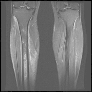
MR images showing marrow oedema from the upper third of the diaphysis to the distal metaphysis of the right tibia. There are also some cortical breaches and localised signal abnormalities consistent with intramedullary abscess (Case 3).
Official websites use .gov
A
.gov website belongs to an official
government organization in the United States.
Secure .gov websites use HTTPS
A lock (
) or https:// means you've safely
connected to the .gov website. Share sensitive
information only on official, secure websites.
MR images showing marrow oedema from the upper third of the diaphysis to the distal metaphysis of the right tibia. There are also some cortical breaches and localised signal abnormalities consistent with intramedullary abscess (Case 3).