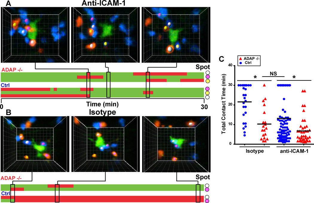
Figure 7. Inhibition of ICAM-1 reduces T cell contact time with antigen-laden DCs.
CTV-labeled wild-type (blue) and CTO-labeled ADAP−/− (red) DO11.10 T cells were co-transferred into CFSE/IFA ear-primed recipients with OVA antigen (Ag). One hour after T cell transfer, 200 µg of anti-ICAM-1 antibody or isotype control antibody was administered by i.p. injection. Draining LNs were harvested 3 hours after antibody treatment and imaged by TPLSM. (A, B) Three-dimensional images and corresponding kymographs of a single DC interacting with transferred T cells in the presence of anti-ICAM-1 antibody (A) or isotype control antibody (B). Colored spots match cell images to contacts on the kymograph. (C) Pooled cumulative T-DC contact time from three separate movies. *, P<0.05 unpaired t test.

