
Figure 5. Sequence conservation at domain interfaces in DnaK-ATP.
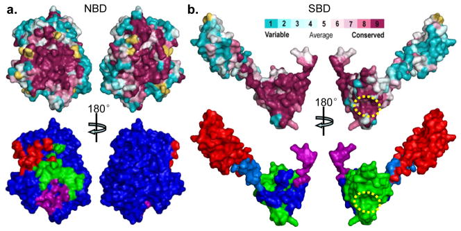
The top panels are sequence conservation at the surface of NBD (a) and SBD (b). Conservation and surface mapping was calculated by ConSurf (http://consurf.tau.ac.il/). The conservation key is shown above b. The dotted yellow circle in b locates the polypeptide-binding site. The bottom panels are mappings of domain interfaces onto the surfaces of NBD (a) and SBD (b). In the bottom panel of a, the surface of NBD is blue, and the imprints of contacts are colored by domain: SBDα (red), SBDβ (green), Linker (purple). In the bottom panel of b, The surfaces of Linker, SBDβ and SBDα are purple, green, and red, respectively, and the imprints of contacts from NBD are colored by the sub-domain receiving that contact: SBDβ (blue), SBDα (marine), and Linker (purple, as it is buried entirely into the interface with NBD).