
Figure 4.
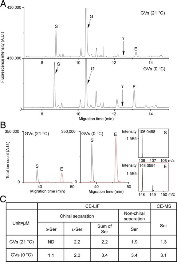
Quantitation of vesicle amino acid content using CE–LIF and CE–MS. To validate quantitation via CE–LIF chiral separations as shown in Figure 3A, two additional measurements using non-chiral separation conditions were performed using CE–LIF and CE–MS detection to validate the quantitative measures and peak assignments. A, Astroglial Sb2-containing vesicles immunoisolated at 21°C and 0°C were analyzed using a non-chiral CE–LIF separation. Arrows point to the serine (S), GABA (γ), glycine (G), and glutamate (E) peaks. B, The same samples were analyzed by CE–MS. Electropherograms were adjusted to the same scale to facilitate comparison between approaches. C, A table shows representative quantification results of serine using these three different methods.