
Figure 1. NKp44+ NK cells are prominently found within MALT.
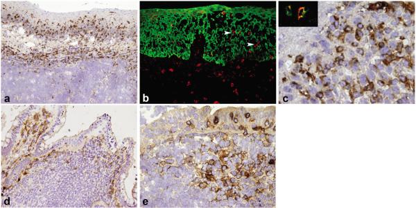
a, Immunohistochemical analysis of tonsil sections with anti-NKp44 shows NKp44+ NK cells residing within the tonsil epithelium and the lamina propria. b, Double immunofluorescence confirms that NKp44+ cells (red) are either in the lamina propria, or within the surface epithelium in close contact with cytokeratin-5+ (green) epithelial cells (white arrowheads). c, At high power view, NKp44+ NK cells show a round to oval morphology and co-express CD56 (inset). d,e, Numerous NKp44+ NK cells are present in the dome region of ileum Peyer's patches (d) and in the luminal epithelium, dome and interfollicular regions of the appendix (e).