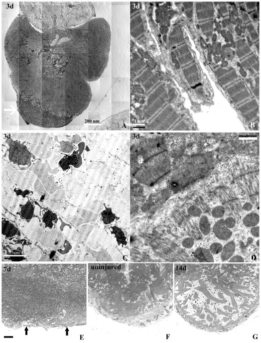
Figure 2.
Characterization of the injury induced by cauterization in plastic sections and by transmission electron microscopy. (A) Montage of a toluidine blue stained 2-um plastic-embedded section of an injured GD ventricle at day 3 post-injury demonstrating a well defined area of injury with loss of myocytes (arrow). (B) Transmission electron micrograph of well organized trabeculated myocytes with well organized sarcomeres, Z-bands, and dense area of mitochondria in a region distal from the injury. (C) Ultrastructure within the injured area showing complete loss of myocyte structure and the presence of crenated nucleated red blood cells contributing to the clot. (D) Ultrastructure of myocytes at the border zone of the injury showing disorganized and lower density of sarcomere closer to the injured area (lower half oh panel), and myocytes with higher sarcomeric density and organization (upper half of panel). (E) Toluidine blue stained section of a heart 7 days after the injury showing a loss of the structural characteristics of the compact and spongy heart showing, and (F) an uninjured heart with well defined compact and the dense trabeculated spongy myocardium. (G) Reconstitution of the compact heart is coupled with the reappearance of a spongy heart of lesser trabecular density at 14 days.

