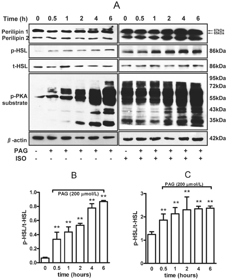
Figure 2. PAG increased phosphorylated PKA substrate, perilipin and hormone sensitive lipase (HSL) in rat adipocytes.
(A) Lysates of adipocytes treated with 200 µmol/L PAG were separated by SDS-PAGE on low-Bis concentration gels and underwent immunoblot analysis with an anti-perilipin antibody. The band shift from 65 kDa (native) to 67 kDa (phosphorylated) perilipin 1 indicates hyperphosphorylation of full-length perilipin 1. The 46-kDa band is perilipin 2. Phosphorylated HSL at Ser659 and phosphorylated PKA (p-PKA) substrate was determined. Relative expression of p-HSL to total HSL was analyzed by band density under basal (B) or isoproterenol (1 µmol/L)-stimulated conditions (C). Data are mean ± SD. ** P<0.01 vs. untreated with PAG.

