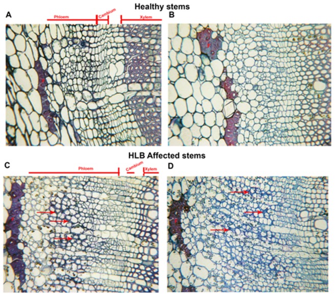
Figure 5. Microscopic analyses of stems of healthy and HLB affected Valencia sweet orange.

A, B. Light microscopy of cross-sections of healthy young stems showing the phloem, cambium and xylem cells. F-Phloem fibers. C,D Cross section of HLB affected young stems showing greater thickness of the phloem layer compared to the healthy. Arrows point to thickened cell walls.
