
Figure 8. Microscopic analyses of roots of healthy and HLB affected Valencia sweet orange on citrumelo rootstock (Citrus paradisi x Poncirus trifoliata).
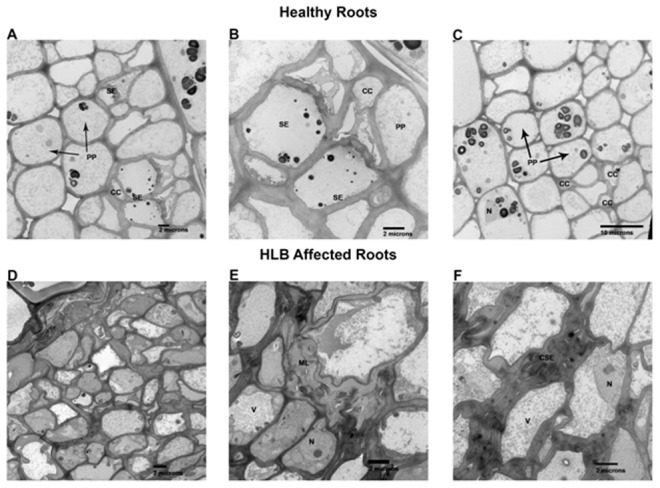
A–D. Electron microscopy of healthy fibrous roots. A & C are relative low magnifications showing size and shape of normal phloem parenchyma (PP), sieve elements (SE) and companion cells (CC). B shows a higher magnification of same areas enhancing the sieve element areas. D–F are HLB affected roots. D is a low magnification comparable to A & C above showing the enriched cytoplasmic contents of these cells compared to the healthy above. E shows enlarged middle lamellas (ML). F shows the collapsed sieve elements (CSE). N, prominent nucleus and V, vacuole.