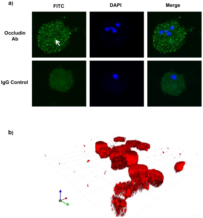
Figure 3. Immunofluorescent confocal microscopy of E. histolytica.
(a) Trophozoites were incubated with or without human C-terminus occludin antibody (Ab) followed by FITC tagged species-specific secondary antibody. DAPI was used to counter stain nuclei. Arrow points to intense green staining for the occludin-like proteins. Note that the intense green staining is absent in the IgG controls. (b) 3D reconstruction of Z-stacks of slices along xy planes of ameba attached on transwell membranes throughout the cellular z-axis at 0.35 µ intervals of trophozoites probed for human occludin C-terminus protein. The red fluorescence tag indicates occludin. An isotype control IgG showed no binding (data not shown). This experiment was repeated twice with similar results.

