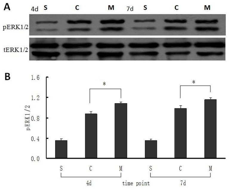
Figure 4. Expression of pERK1/2 in intestinal mucosa at 4 and 7 days after MSCs transplantation.
(A) Bands of the expression of pERK1/2 and its own tERk1/2 protein were detected by western blot. (B) The ratio value of pERK1/2 to its own tERK1/2 of the bands which was evaluated densitometrically using the software Quantity One for the three groups at different time points. Each bar represents mean ± SD of the ratio value in every group. S: sham group; C: control (saline) group; M: MSCs group. 4d and 7d represent 4 days and 7 days postoperatively. *: P<0.05, compared with control (saline) group, by one-way ANOVA test.

