
Figure 1. Increased CD11cHIMHCIIHI DCs in H. pylori–infected mouse stomach and the expression of TLRs by H. pylori-stimulated BMDCs.
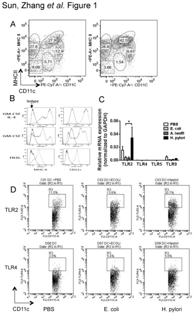
a, Single cell prep of stomach from uninfected C57BL/6 mice or from mice infected with H. pylori for 24 h were dual-labeled with phycoerythrin (PE)-conjugated anti-mouse MHC II and PE and a cyanine dye (Cy7)-conjugated anti-mouse CD11c antibodies and analyzed by FACS. b, BMDCs from C57BL/6 mice were cultured in culture medium supplemented with GM-CSF plus IL-4, GM-CSF only, or Flt3L.The expression of MHC II and CD11c was analyzed by flow cytometry on day 6. Background staining was assessed using isotype controls. BMDCs in the GM-CSF plus IL-4 group had higher levels of MHC II and CD11c than cells in the GM-CSF group or Flt3L group. c, BMDCs were pulsed with PBS, live Escherichia coli (EC), live Acinetobacter lwoffii (AL), or live H. pylori (HP) for 18 h (multiplicity of infection, 10:1). The mRNA expressions of TLR 2, TLR4, TLR5, and TLR9 were measured by quantitative PCR. Data shown were obtained from three independent experiments. d, BMDCs were pulsed with PBS, live Escherichia coli (EC), or live H. pylori (HP) for 18 h (multiplicity of infection, 10:1). BMDCs were dual-labeled with fluorescence-conjugated CD11c and TLR2 or TLR4 and analyzed by flow cytometry. Representative dot plots from three separate experiments are shown.