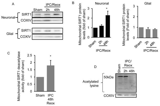
Figure 2. IPC increases mitochondrial SIRT1 protein levels specifically in neurons.
Primary cortical glial and neuronal-only cultures were exposed to oxygen and glucose deprivation for 45 min (IPC) and the level of mitochondrial SIRT1 was determined 2 and 48 hrs later (n = 6) (A). Western blot quantitation is shown in (B). SIRT1-specific deacetylase activity was determined in mitochondrial lysates from neuronal cultures 48 hrs following exposure to sham or IPC (n = 4) (C). Neuronal mitochondria acetylation levels were determined at 2 and 48 hrs following IPC exposure (n =3) (D). * p < 0.05 increase from sham by one-way analysis of variance (ANOVA) and Dunnetts’ multiple comparison post hoc test.

