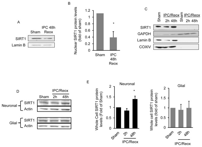
Figure 3. IPC increases SIRT1 expression and decreases nuclear SIRT1 protein levels.
Cortical neuronal cultures were exposed to oxygen and glucose deprivation for 45 min (IPC) and nuclear SIRT1 protein levels determined 48 hrs later (n = 5) (A). Western blot quantitation of nuclear SIRT1 protein levels is shown in (B). The effect of IPC on cytoplasmic SIRT1 protein levels is shown in (C). The soluble cytoplasmic fraction was isolated from the nuclear and mitochondrial fractions as described in methods. The purity of the cytoplasmic fraction was determined by reprobing the blot for the cytoplasmic marker, GAPDH, with the nuclear marker lamin B and with the mitochondrial marker COXIV. In (D) whole cell SIRT1 protein levels in cortical glial and neuronal only cultures are shown at 2 and 48 hrs following IPC exposure (n=8). Western blot quantitation is shown in (E). Data are means ± SEM compared to sham treated animals. * p < 0.05 increase from sham by one-way analysis of variance (ANOVA) and Dunnetts’ multiple comparison post hoc test.

