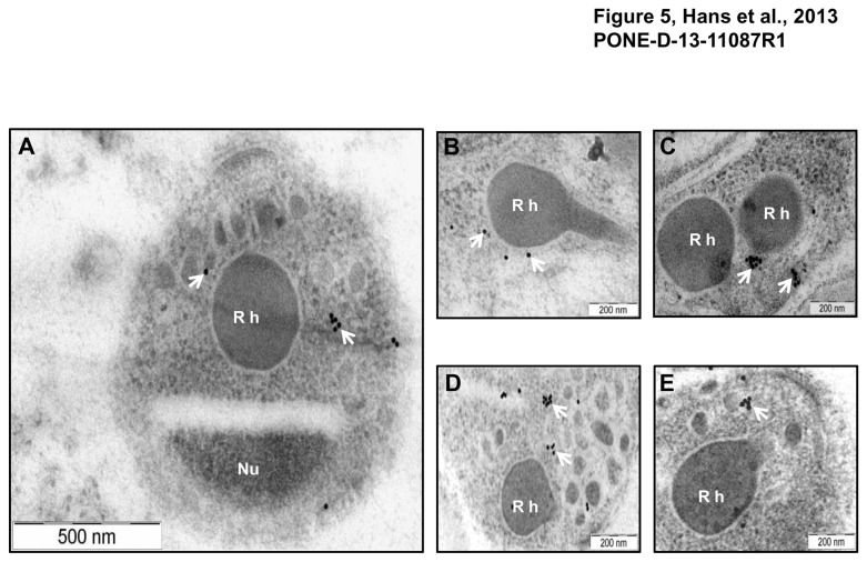
Figure 5. Localization of PfMA by Immuno-electron microscopy.
Ultra thin sections of P. falciparum parasites at schizont/ merozoite stages were labelled with mouse anti-PfMA or anti-EBA-175 antibodies followed with anti-mouse secondary antibody conjugated with 15nm colloidal gold particle. (A) Labelling with the PfMA antibody was confined only to the micronemes (arrows) in the apical end of the free merozoite and was absent from the rhoptry (Rh) or nucleus (Nu). (B, C) Similarly PfMA staining in the schizonts was observed only in the micronemes and not in the rhoptries. (D, E) A similar pattern of staining in the schizonts was observed with the micronemal marker EBA-175.

