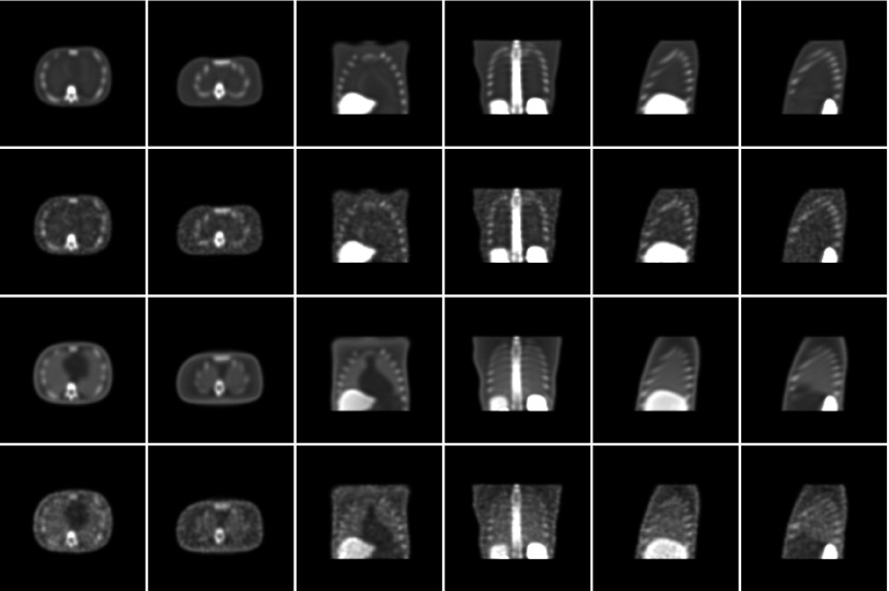
Figure 2.
Comparison of transverse, coronal, and sagittal images extracted from normal reconstructions. Each column pertains to a fixed slice from the NCAT phantom. From top to bottom, the rows represent: (1) noise-free AllC reconstructions; (2) noisy AllC reconstructions; (3) noise-free RC reconstructions; and (4) noisy RC reconstructions.

