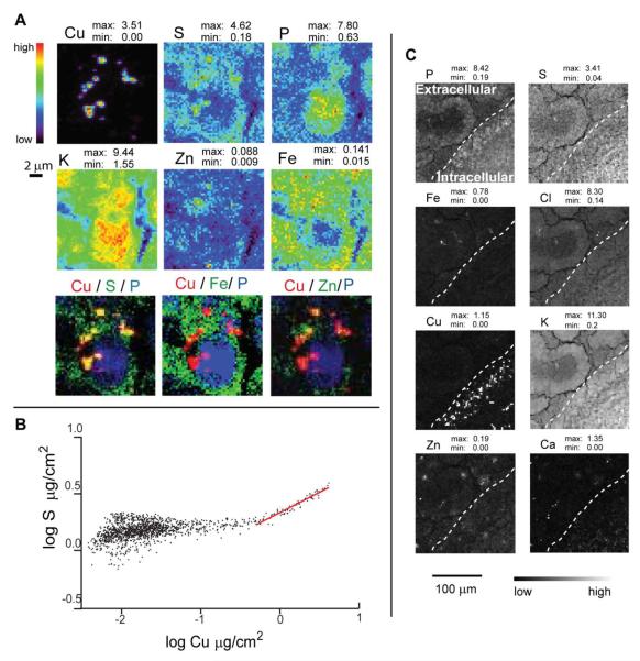
Figure 2.
A. XRF image of the SVZ. Elemental concentrations are in μg / cm2 in 10 μm thick sections; experimental details are given in Table 1. The dashed line shows the ventricle wall with brain tissue in the right low corner. From XRF imaging it is evident that SVZ selectively accumulates Cu over other trace elements. B. A linear fit of the Cu/S correlation allows estimation of the S/Cu molar ratio (Table 3) suggesting that the coordination of Cu is accomplished by sulfur ligands. C. XRF microscopy clearly demonstrates intracellular structures with dramatically increased Cu content. The cell nucleus is identified by the increased phosphorous signal, while sulfur is distributed throughout the cytoplasm. Potassium has much higher concentration inside the cell than outside and, thus, helps to define the overall cell shape. The three-color plots in Fig. A show that Cu co-localizes with S. Elemental contents are given in μg/cm2.

