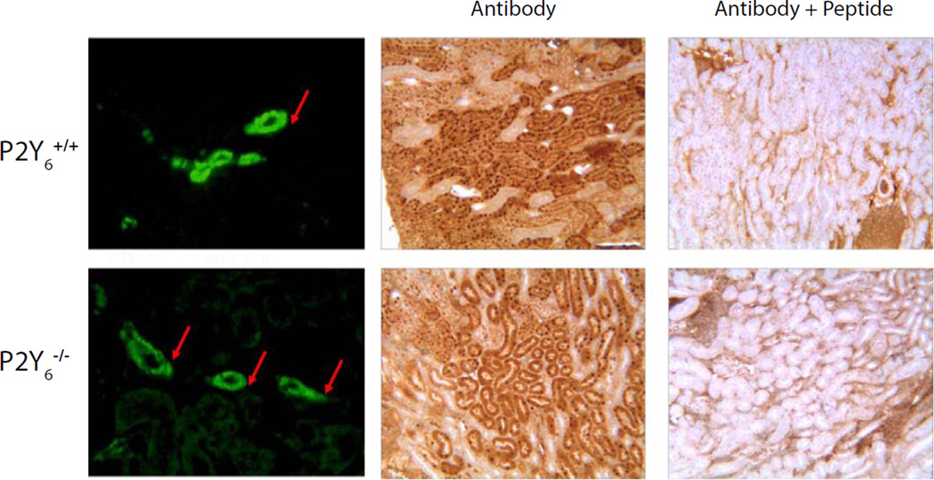
Figure 3.
Immunostaining of kidney sections from control and knockout mice using APR-011. Left panels show immunofluorescent labeling of tubules (arrows) in both P2Y6 positive and negative tissue. Right panels show peptide competition of the antibody resulting in dramatically diminished binding to kidney sections from both control and KO animals (middle panels).

