
Figure 1.
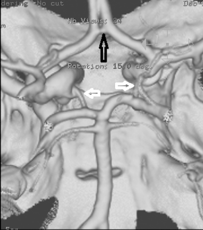
Circle of Willis anatomy: Surface volume rendered image from a CT angiography (CTA) showing the anterior communicating artery (open black arrow) and bilateral posterior communicating arteries (open white arrows). Orientation is as though the brain has been removed and we are looking down at the inside of the skull from above.