
Figure 1.
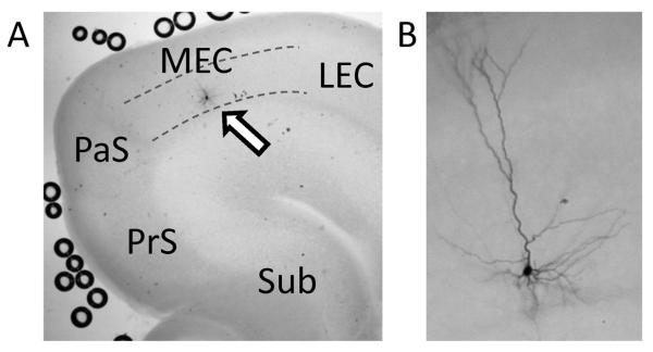
Location and morphology of MEC layer III neurons from which recordings were obtained. (A) A low magnification image showing a biocytin stained MEC layer III neuron (arrow). Dotted lines indicate the upper and the lower borders of the layer III. Sub: subiculum, PrS: presubiculum, PaS: parasubiculum, LEC: lateral entorhinal cortex. (B) A higher magnification image showing another example of biocytin stained MEC layer III neuron. Typical pyramidal cell morphology can be seen.