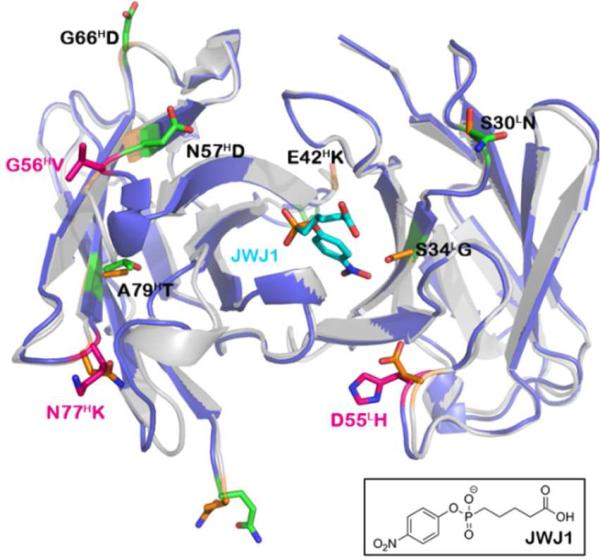
Figure 1.
Alignment of germline 48G7g structure (gray, PDB code: 1AJ7) with matured 48G7 (blue, PDB code: 1GAF). Somatic mutations involved in binding are highlighted in pink (G56HV, N77HK, and D55LH). The remaining mutations are denoted in green (E1HQ, E42HK, N57HD, G66HD, A79HT, S30LN, and S34LG). The corresponding residues in the germline structure are denoted in orange. JWJ1 is shown in its high-affinity binding conformation (cyan) with its chemical structure included below.

