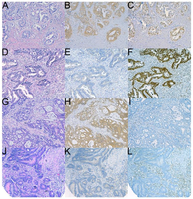
Figure 2.
Serial sections of four different CRCs stained with hematoxylin and eosin (A,D,G,J), and IHC for BRAFV600E (B,E,H,K) and MLH1 (C,F,I,L). The tumor in A-B-C is MSS (positive staining was also seen for PMS2, MSH2 and MSH6) and BRAFV600E positive. This appears to represent a poor prognostic group. The tumour in D-E-F is BRAFV600E negative and MSS. The tumor illustrated in G-H-I shows negative staining for MLH1 and demonstrates MSI. It also demonstrates positive staining for BRAFV600E therefore LS is essentially excluded. The tumor illustrated in J-K-L demonstrates MSI and negative staining for BRAFV600E therefore in this instance formal genetic testing for LS is justified. As illustrated in panels B and H, positive staining for BRAFV600E is characterized by widespread cytoplasmic staining limited to tumor cells (Original magnifications 100X).

