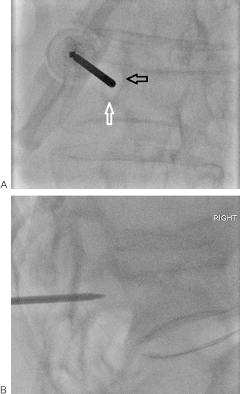
Figure 3.

(A, B) Ipsilateral oblique and lateral fluoroscopic views of a T10 vertebral fracture. The needle should be advanced at the upper and outer quadrant of the pedicle. The needle must be advanced without violating the inferior (white arrow) and medial (black arrow) walls of the pedicle to avoid potential nerve or cord injury. Intermittent lateral views should be obtained to ensure appropriate angle of entry into the vertebral body.
