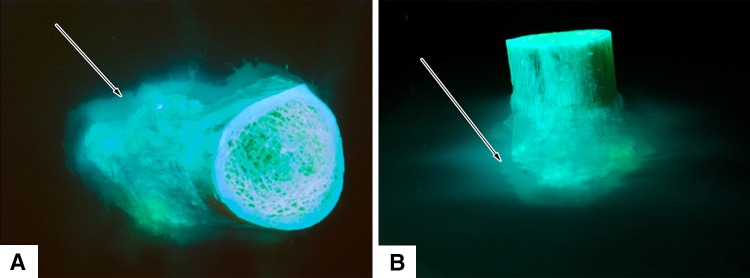
Fig. 5A–B .

A distal femoral metaphysis loaded with doxycycline for visualization of fluorescence at the periosteal surface is shown in (A) axial and (B) transverse views. Doxycycline egress is seen as bright green fluorescence. Arrows indicate a cloud of fluorescence that has diffused into the agarose gel and become confluent from focal areas of doxycycline released from vascular channels.
