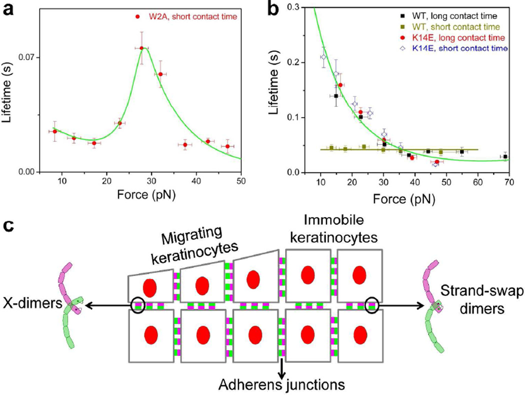
Figure 2. Mechanical force tunes the kinetics of cadherin adhesion.
Adapted from (Rakshit et al., 2012). (A) X-dimers form catch bonds which become longer lived and lock in the presence of tensile force. When W2A cadherin X-dimers are pulled, their bond lifetimes increase with force. After reaching a maximum at a critical force of ~ 30 pN, the lifetimes decrease. (B) Strand swap dimers form slip bonds which become shorter lived when pulled. Slip bonds are formed by K14E mutants that interact for short and long periods of time and also by WT cadherins that interact for long periods of time. However, when WT cadherins interact for a short period of time, they form ideal bonds that are insensitive to force. (C) Hypothetical mechanism by which keratinocytes resist tensile forces during skin renewal and wound healing. As skin cells reposition themselves, E-cadherins bind rapidly to form X-dimers that allow cells to grip strongly under load. In immobile keratinocytes, E-cadherins form more robust strand-swap dimers that have a high affinity in the absence of force.

