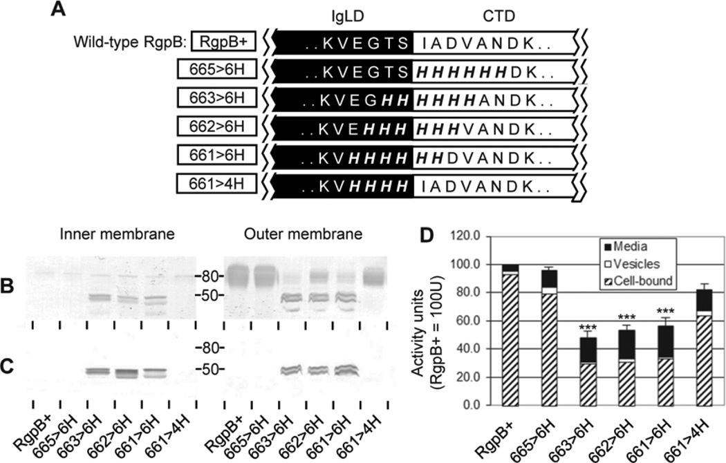
Figure 4. Hexa-histidine scanning mutagenesis of the IgLD-CTD junction.
(A) Various mutants (boxed) with 4 or 6 consecutive junctional residues changed to 4×His or 6×His, respectively. Early stationary phase cultures of each mutant were normalised to OD600 1.5 and subjected to inner membrane and outer membrane fractionation as per Methods. 50 µL of each membrane fraction was analyzed by Western blot using (B) anti-RgpB and (C) anti-CTDRgpB to determine the maturation status and partitioning of RgpB. (D) 20 µL of normalized cultures were assayed for media, vesicle-bound and cell-associated RgpB activity using synthetic substrates as per Methods. Data represents the mean and SEM from four independent experiments with the total activity in the control RgpB+ mutant equaling 100 U. Significance of total Rgp activity vs. RgpB+ wild-type control: ***, p<0.001.

