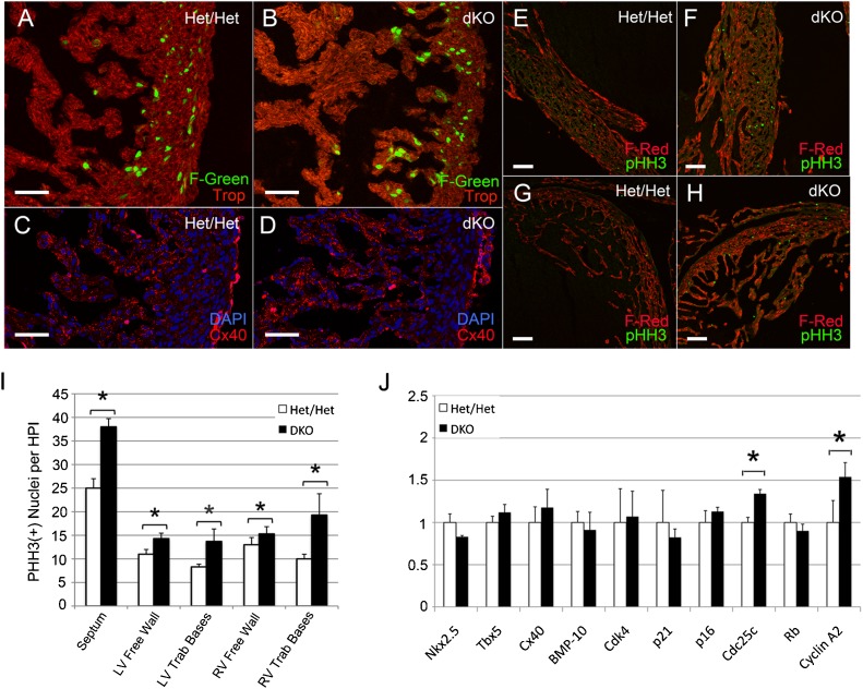
Fig. 6. Analysis of dKO/Fucci-Green (dKO/F-Green) and dKO/Fucci-Red (dKO/F-Red) hearts (A–H).
(B) Non-compacted E14.5 dKO/F-Green hearts remained committed to trabecular expansion and eccentric remodeling as evidenced by persistent F-Green positive nuclei at trabecular bases. (D) The hypertrabecularized region was composed of Cx40 positive myocytes. (E–H) dKO/F-Red hearts at E15.5 were immunostained for pHH3 to quantify the number of cells undergoing mitosis. (F,H) dKO hearts exhibited a significant increase in mitotic cells throughout the compact myocardium and in the trabecular bases. (I) The number of mitotic cells in the interventricular septum, LV and RV compact myocardium, and trabecular bases was quantified per high power field (*P<0.05). (J) Semi-quantitative RT-PCR analysis of E14.5 dKO ventricles vs Het/Het littermate controls. Both Cyclin A and CDC25 were significantly higher in dKO ventricles (*P<0.05). Scale bars: 50 µm (A–D), 100 µm (E–H).

