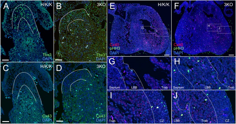
Fig. 9. 3KO hearts maintain appropriate localization of proximal VCS and trabecular markers.
(A,C) In H/K/K littermates, Tbx3 was localized to the proximal VCS, where it repressed Cx43 expression. (B,D) The reciprocal pattern of Tbx3 and Cx43 expression was maintained in the 3KO proximal VCS. (E,F) In comparison to littermate hearts, 3KO ventricular chambers were filled with two populations of Cx40 positive myocytes. (G) The left bundle branch (LBB) in control hearts was identified as a thin band of Cx40 low-expressing myocytes adjacent to the septum. (G,I) The remaining trabecular myocardium (Trab) was composed of Cx40 high-expressing myocytes. (H,J) 3KO ventricles showed marked expansion of Cx40 low expressing, left bundle branch (LBB) myocytes and lateralization of Cx40 high-expressing trabecular myocytes (Trab) within the LV chamber. (I,J) The boundary between the trabecular region (Trab) and the compact myocardium (CZ) remains intact in 3KO hearts. (Troponin, Trop; Connexin-40, Cx40; Connexin-43, Cx43; Nuclear stain, DAPI). Scale bars: 50 µm (A–D), 100 µm (E,F).

