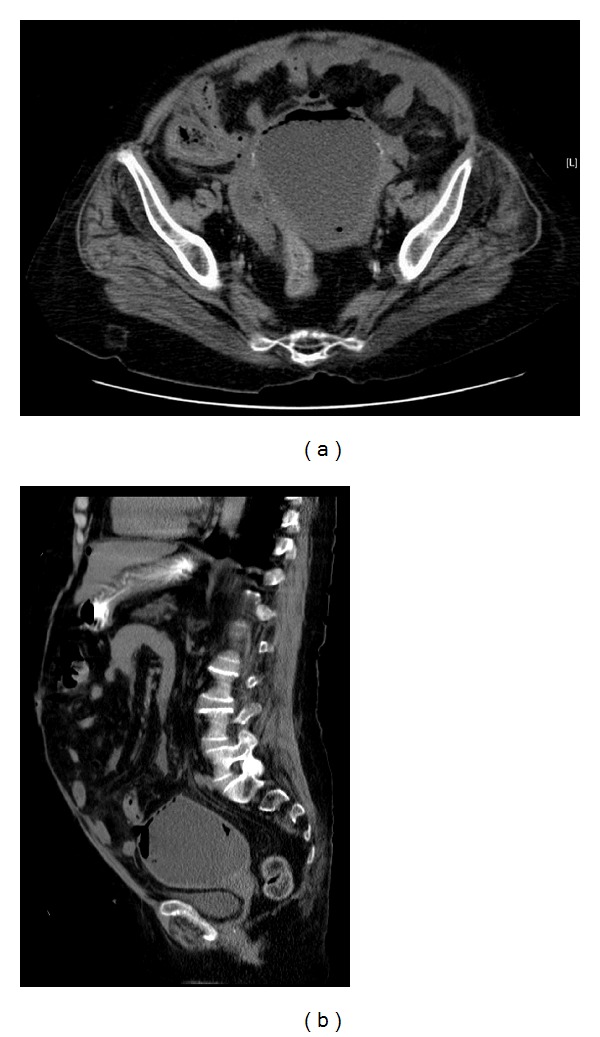
Figure 2.

Transverse (a) and sagittal (b) views of noncontrasted-enhanced computed tomography (CT) scan showing a significantly distended fluid-filled uterus measuring 10 × 7.8 × 10 cm, in addition to a single focus of perforation involving the uterine fundus and associated with presence of free air within the nondependant area. No evidence of ascites or pelvi-abdominal lymphadenopathy was identified.
