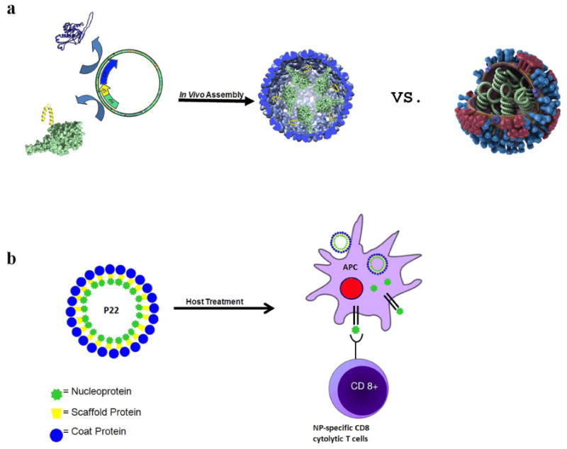
Figure 1. Schematic representations of the expression and in vivo encapsulation of the nucleoprotein through programmed self-assembly of the P22 VLP and the biomimetic display in order to elicit nucleoprotein-specific CD8+T cell response.

a) Nucleoprotein (NP; green) fusion with the scaffold protein (SP; yellow) is co-expressed with the coat protein (blue), resulting in assembly of the NP-P22 VLP. A model of the natural influenza virus (made available by the Center for Disease Control) is shown illustrating the display of NP (green), neuraminidase (red), hemagglutinin (blue), and M2 ion channels (purple) to highlight the biomimetic design of the NP-P22. b) Treatment of a host (immunization) with NP-P22, due to its biomimetic display of NP, is expected to be processed by the pathway that generates CD8+ T cells specific for NP.
